
Actual cases treated by Dr. Mortensen
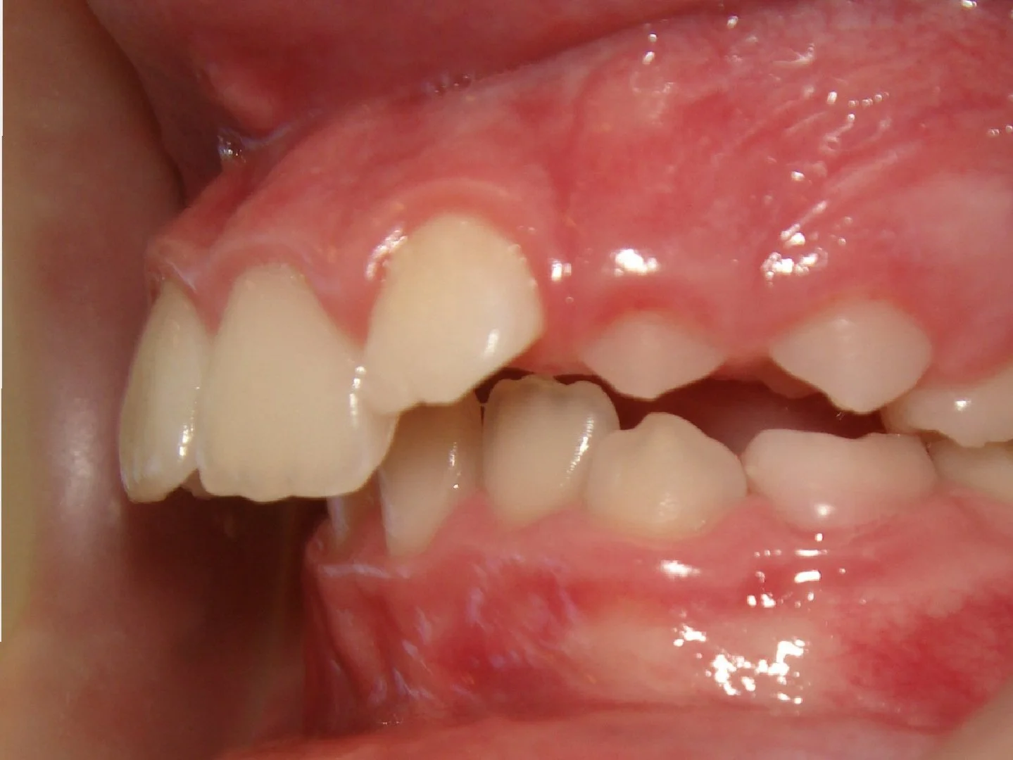
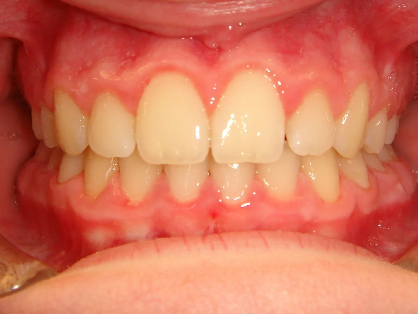
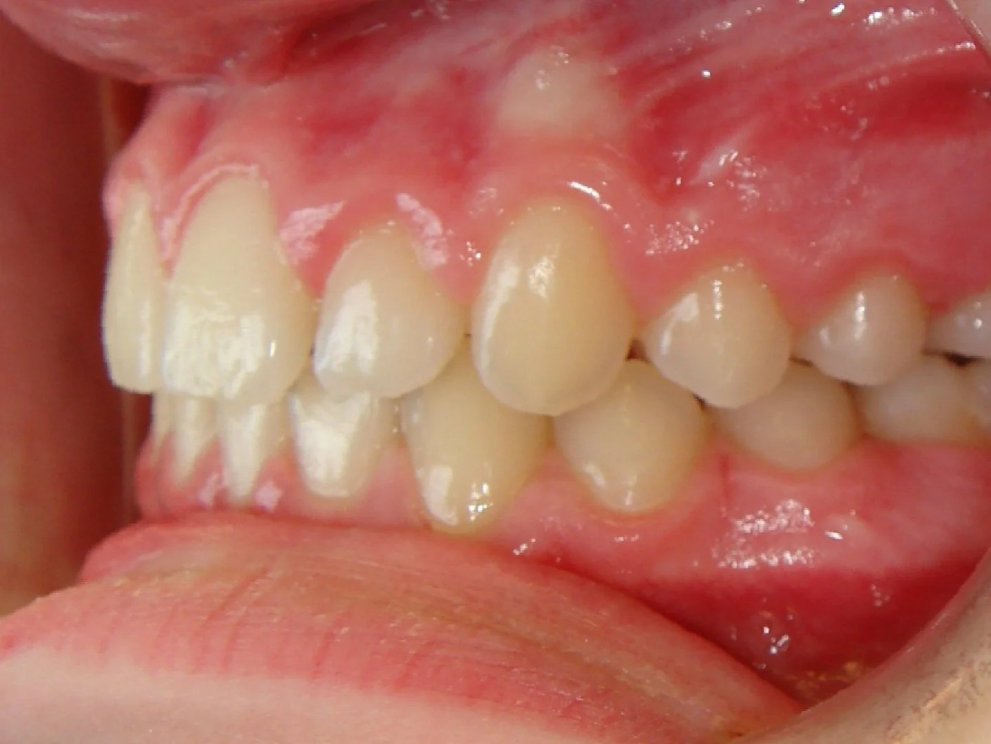
Case #1: Excess Overjet / Overbite & Crowding

Before

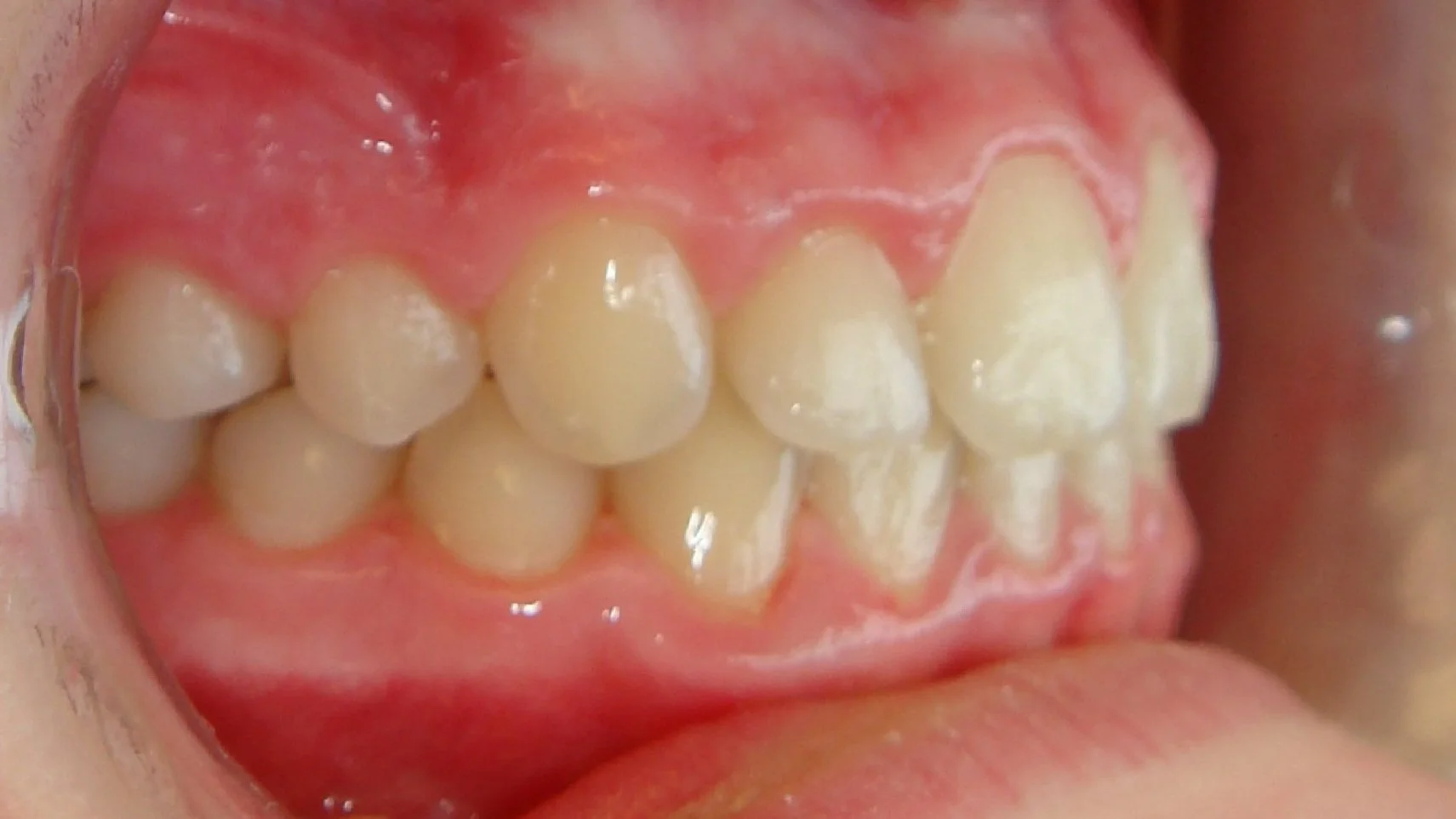
After
-
Chief Concern
“The top teeth stick out too far”.
This 10 year old girl had crowding of her upper and lower teeth, and an extreme overjet and overbite. Her case was treated over 2 years with braces and elastics.
Case #2: Adult Underbite

Before: Severe Underbite


After: Perfect Profile

After: Perfect Bite

-
Chief Concern
“I want to fix my underbite. I need it done in under a year if possible!”
Considering that the average case of non-surgical braces takes about 2 years, this was a significant task! However, we finished in 12 months with beautiful results. Her treatment included full braces combined with orthognathic (jaw) surgery.
Case #3: Phase I, Impacted Canines
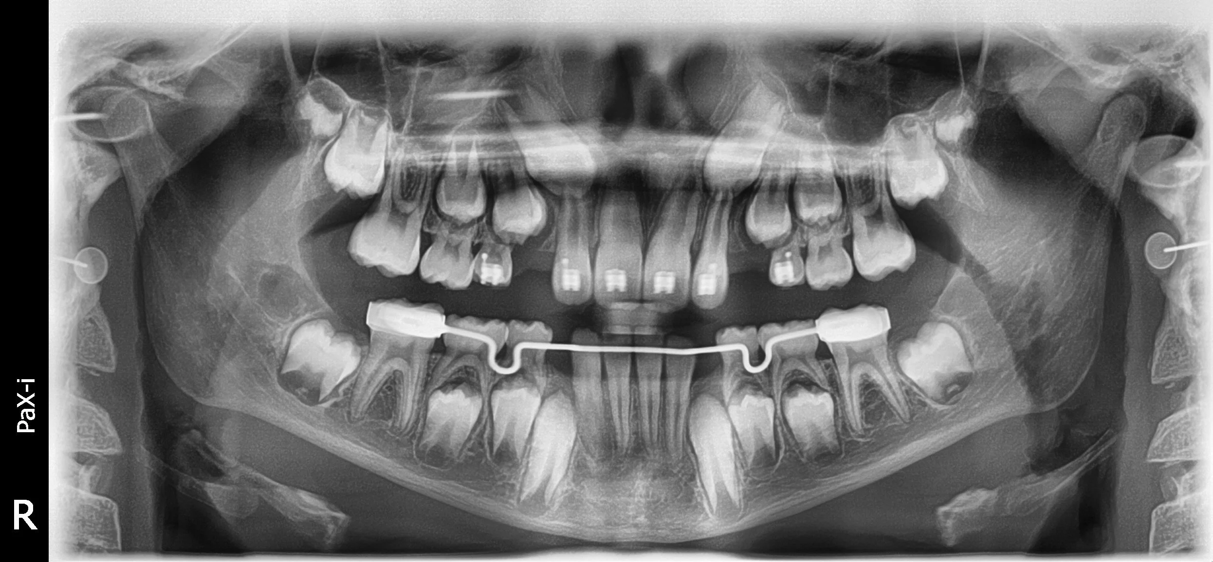
Start

Progress

Finish
-
Chief Concern
Patient was referred by her pediatric dentist for evaluation of upper canines.
Both upper canines were significantly impacted and risked damaging the permanent lateral incisors. Braces were placed as part of a Phase I (early) treatment in order to save the lateral incisors and bring the canines into position. Small chains were attached to the canines and they were gently brought into the arch over 18 months.